AI‑Powered Handheld Microscope Expands Depth of Field for Early Cancer Detection
PrecisionView delivers eight times the depth of field and five times the field of view of conventional microscopes while keeping cellular resolution, aiming to enable real‑time, biopsy‑free cancer screening in low‑resource settings.
PrecisionView, an AI‑powered handheld microscope, expands depth of field eightfold and field of view fivefold while preserving cellular resolution, allowing real‑time visualization of both nuclear and vascular tissue features. Priced at roughly $3,000, the device aims to bring biopsy‑free cancer screening to clinics and low‑resource settings.
Researchers at Rice University and MD Anderson Cancer Center designed PrecisionView to solve a long‑standing limitation of endoscopic microscopy: the trade‑off between seeing fine cellular detail and covering a large tissue area. Traditional systems force clinicians to pick either high resolution over a small field or a broader view with blurred depth. By integrating a custom phase mask with a deep‑learning reconstruction algorithm, the team redesigned the optics themselves rather than relying on post‑processing AI.
The researchers say PrecisionView eliminates the need to choose between image detail and coverage, allowing clinicians to see both clearly in real time. Quantitatively, PrecisionView provides about five times the field of view and eight times the depth of field of conventional microscopes while maintaining cellular‑level resolution. Practically, the microscope is assembled from inexpensive, off‑the‑shelf components and costs approximately $3,000. This low price point enables deployment in community clinics, mobile health units, and settings where traditional pathology infrastructure is lacking.
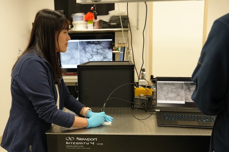
In validation experiments, the team scanned the oral cavities of healthy volunteers, producing high‑resolution maps of tissue architecture and vasculature across areas larger than one square centimeter at up to 15 frames per second. The continuous view let them trace blood vessel patterns that would be missed with a narrow‑field scope. Separately, the microscope was applied to cervical tissue samples containing precancerous lesions. It clearly distinguished abnormal nuclear patterns from surrounding healthy tissue, demonstrating that the combined cellular and vascular contrast can flag early neoplastic change without a biopsy. These results suggest that PrecisionView could reduce false‑negative rates by sampling a larger tissue area in real time, potentially cutting down on unnecessary follow‑up procedures. For clinicians, the handheld form factor means the device can be maneuvered easily over irregular surfaces, such as the inner cheek or cervix, without losing focus. Practical takeaways for readers include: a) the tool offers a non‑invasive, real‑time alternative to biopsy for initial cancer screening; b) its cost is low enough for widespread adoption in low‑ and middle‑income countries; c) the AI‑optimized optics approach may be adapted to other endoscopic or surgical imaging systems needing greater depth of field.
Next steps involve multicenter clinical trials to measure how PrecisionView affects early detection rates and to evaluate its integration with digital pathology platforms for remote expert review.
Continue reading
More in this thread
AI‑Designed Handheld Microscope Expands Field of View and Depth for Real‑Time Cancer Detection
Dr. Priya Sharma
WHO Says Hantavirus Outbreak on Cruise Ship Is Not Another COVID‑19
Dr. Priya Sharma
Spain Confirms Hantavirus Case in Cruise Ship Evacuee as WHO Reports 11 Infections, Three Deaths
Dr. Priya Sharma
Conversation
Reader notes
Loading comments...