AI‑Designed Handheld Microscope Expands Field of View and Depth for Real‑Time Cancer Detection
The AI‑powered PrecisionView microscope offers five‑fold wider view and eight‑fold greater depth than standard devices, costs about $3,000, and could enable real‑time cancer screening in low‑resource settings.
TL;DR
A handheld microscope named PrecisionView uses AI‑optimized optics to deliver five times the field of view and eight times the depth of conventional devices while preserving cellular detail, all for roughly $3,000.
Context Early detection markedly improves survival for epithelial cancers such as cervical and oral carcinoma, yet current microscopes force a trade‑off between seeing fine detail and covering a large area. This limitation often leads to missed lesions or the need for invasive biopsies, especially in low‑resource settings where pathology labs are scarce. In Nigeria, cervical and oral cancers contribute significantly to cancer mortality, highlighting the need for affordable point‑of‑care tools.
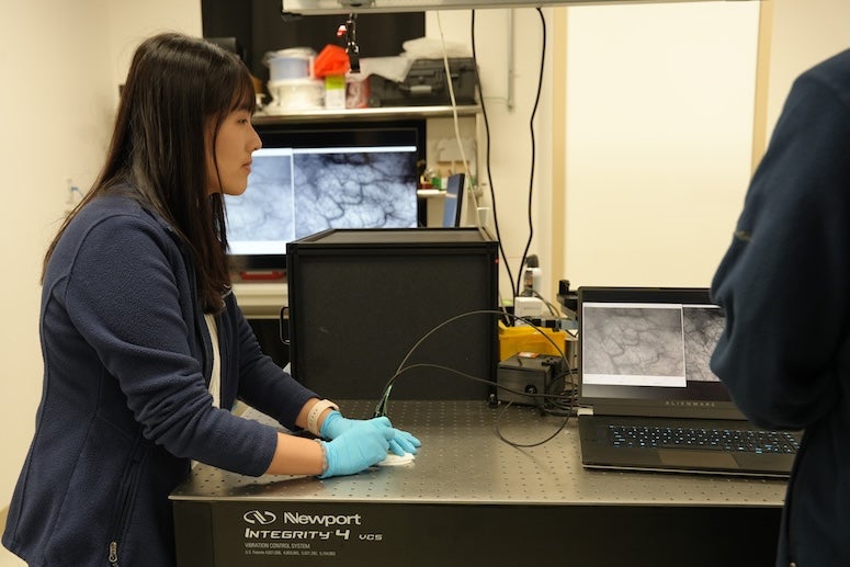
Key Facts - PrecisionView provides about five times the field of view and eight times the depth of field of standard in‑vivo microscopes while maintaining subcellular resolution (Fact 2). - The system’s core is a custom phase mask designed by a deep‑learning algorithm; the AI‑engineered optics reshape light before detection, and a reconstruction algorithm restores clear images in real time. - Built from readily available components, the device costs approximately $3,000, making it feasible for clinics, mobile units, and low‑resource hospitals (Fact 3). - Imaging speeds reach up to 15 frames per second, allowing clinicians to view nuclear alterations and underlying microvascular patterns across tissue areas of several square centimeters without pausing for focus adjustments. - In a pilot study, researchers scanned the oral cavity of healthy volunteers and examined cervical tissue containing precancerous lesions. The device distinguished abnormal regions from healthy tissue, though the study was not a randomized controlled trial and exact sample sizes were not reported.
What It Means By removing the detail‑coverage trade‑off, PrecisionView could enable clinicians to screen larger lesions in a single glance, reducing the chance of missing early cancer signs. Its low price and portability suggest a role in community screening programs and in settings where sending tissue to a central lab causes delays. However, the technology remains at the proof‑of‑concept stage; prospective studies with larger cohorts are needed to confirm diagnostic accuracy, assess operator learning curves, and determine whether same‑day imaging can cut biopsy rates and improve patient outcomes. Regulatory clearance and scalable manufacturing will be key next steps.
Watch for upcoming multicenter trials that will compare PrecisionView’s sensitivity and specificity against standard histopathology, as well as efforts to pursue FDA or CE marking and to integrate the device with telepathology networks for remote expert review.
Continue reading
More in this thread
Conversation
Reader notes
Loading comments...